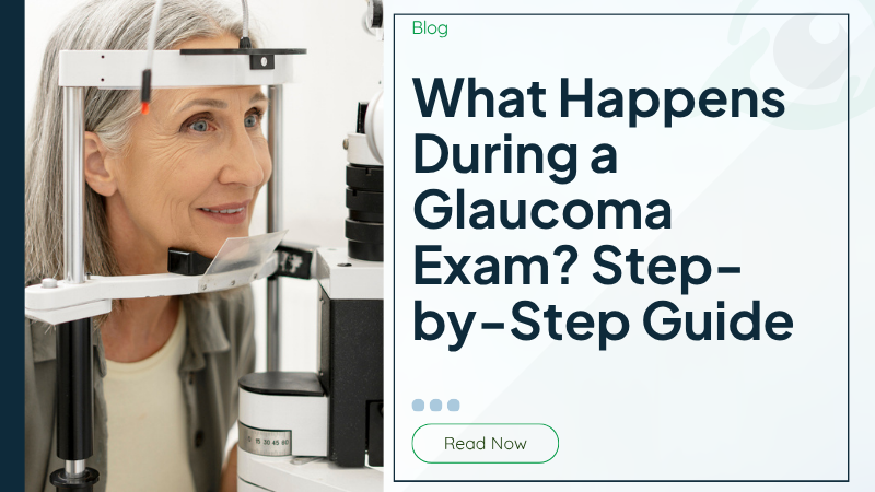
A glaucoma exam begins with a review of your medical history and any symptoms you may be experiencing. Your eye doctor will ask about your general health, medications, family history of glaucoma, previous eye conditions, and whether you've noticed any changes in your vision. Since glaucoma often runs in families, this information helps determine your individual risk level.
Next, your visual acuity is checked using a standard eye chart. Although glaucoma usually affects peripheral vision before central vision, measuring your overall eyesight establishes an important baseline for future comparisons.
One of the most important parts of the examination is measuring your intraocular pressure using a test called tonometry. Elevated eye pressure is a major risk factor for glaucoma, although some people develop glaucoma even when their eye pressure falls within the normal range. Tonometry is quick, painless, and takes only a few moments for each eye.
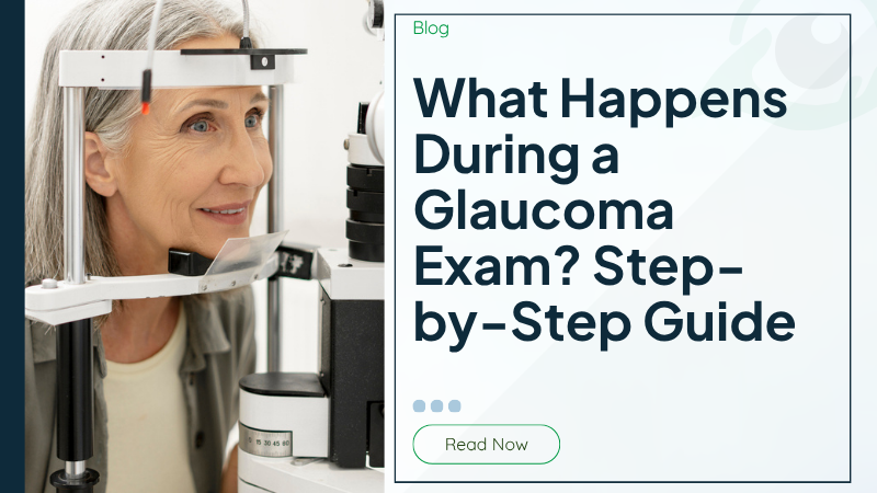
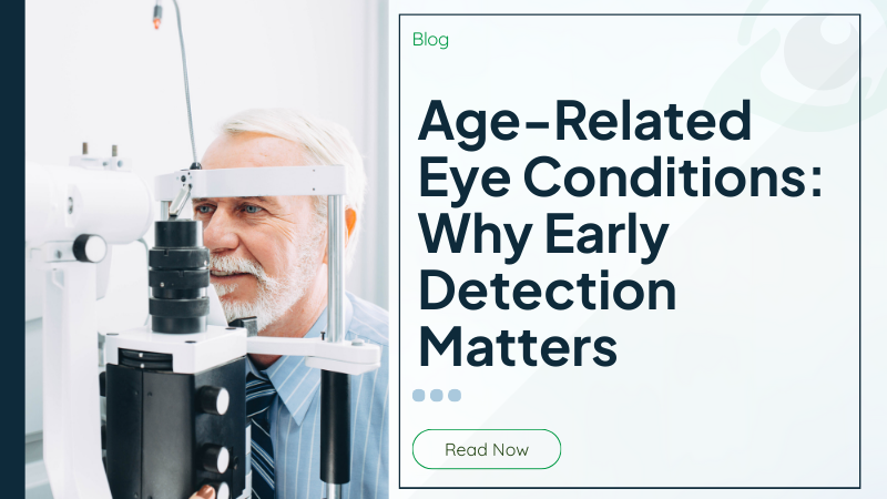
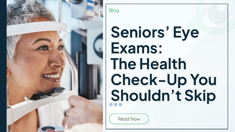
Your eye doctor will then perform a slit lamp examination. This specialized microscope provides a highly magnified view of the front structures of the eye, including the cornea, iris, and lens. It also allows the doctor to assess the overall health of your eyes before continuing with additional testing.
The optic nerve is carefully examined because glaucoma damages the nerve fibers responsible for transmitting visual information from the eye to the brain. Healthy optic nerves have a characteristic appearance, while glaucoma may cause subtle structural changes that can often be detected before vision is affected.
Your pupils may also be dilated to provide a wider view of the optic nerve and retina. Although dilation temporarily causes blurred near vision and increased sensitivity to light, it allows your doctor to perform a more comprehensive examination of the back of the eye.
A typical glaucoma exam may include:
- Medical and family history review
- Vision assessment
- Eye pressure measurement (tonometry)
- Slit lamp examination
- Optic nerve evaluation
- Pupil dilation
- Retinal examination
- Comprehensive eye health assessment
Each step contributes valuable information. Rather than making a diagnosis based on one result alone, your eye doctor evaluates the findings together to determine whether glaucoma is present or whether additional testing is recommended.
Even if everything appears normal, these baseline measurements become extremely valuable for future appointments. Comparing today's results with future examinations helps identify even subtle changes that may develop over time.
GLAUCOMA TESTING
If your examination identifies potential risk factors or suspicious findings, your eye doctor may recommend additional glaucoma testing. These specialized evaluations provide detailed information that helps confirm a diagnosis, establish a baseline, and monitor changes over time.
One of the most common tests is a visual field assessment, also known as perimetry. During this test, you look straight ahead into a specialized machine while pressing a button whenever you see small lights appear in your peripheral vision. Because glaucoma often affects side vision first, this examination helps detect areas of vision loss that you may not notice during everyday activities.
Another important test is Optical Coherence Tomography (OCT). OCT uses light waves to create highly detailed cross-sectional images of the retina and optic nerve. This painless imaging technology allows your eye doctor to measure the thickness of the retinal nerve fiber layer and detect microscopic changes long before vision loss becomes noticeable.
Some patients also undergo pachymetry, which measures the thickness of the cornea. Corneal thickness affects eye pressure readings, so this measurement helps your doctor interpret tonometry results more accurately and assess your overall glaucoma risk.
Gonioscopy is another specialized examination that evaluates the drainage angle inside the eye. This test helps determine whether the drainage angle is open or narrow, allowing your eye doctor to distinguish between different types of glaucoma and recommend the most appropriate treatment plan.
Retinal photography may also be performed to create permanent images of the optic nerve. These photographs allow your doctor to compare changes over time and monitor disease progression during future visits.
Specialized glaucoma testing may include:
- Visual field testing
- Optical Coherence Tomography (OCT)
- Pachymetry
- Gonioscopy
- Retinal photography
- Optic nerve imaging
- Repeat eye pressure measurements
- Baseline vision documentation
Most of these tests are completely painless and require only a few minutes to perform. Depending on your individual circumstances, not every test is necessary during every appointment. Your eye doctor selects the examinations that provide the most useful information based on your eye health and risk factors.
Technology has greatly improved the ability to detect glaucoma at its earliest stages. Advanced imaging allows doctors to identify structural changes before noticeable vision loss develops, making early treatment far more effective.
Another important benefit of comprehensive glaucoma testing is the ability to establish a baseline for your eye health. Even if no signs of glaucoma are found during your first examination, the information collected becomes an important reference for future visits. Comparing eye pressure measurements, optic nerve images, and visual field results over time allows your eye doctor to identify even the smallest changes that could indicate the early development of glaucoma. Detecting these subtle differences before symptoms appear significantly improves the chances of preserving your vision.
Regular follow-up appointments are equally important because glaucoma is a chronic condition that can develop gradually over many years. Some individuals with elevated eye pressure never develop glaucoma, while others with normal eye pressure may still experience optic nerve damage. For this reason, your eye doctor evaluates all test results together rather than relying on a single measurement. By combining advanced diagnostic technology with routine monitoring and personalized care, glaucoma exams provide one of the most effective ways to detect problems early, guide treatment decisions, and protect your long-term eye health and quality of life.
AFTER YOUR EXAM
Once all testing has been completed, your eye doctor reviews the results with you and explains what they mean. Many patients receive reassuring news that their eyes are healthy and show no signs of glaucoma. Others may learn that they have certain risk factors requiring closer monitoring, while some patients may receive a diagnosis that allows treatment to begin before significant vision loss occurs.
If glaucoma is detected, treatment often begins with prescription eye drops designed to lower eye pressure. Depending on the type and severity of glaucoma, laser treatment or surgery may also be recommended. Early treatment plays a critical role in slowing disease progression and preserving long-term vision.
If your examination does not reveal glaucoma but you have increased risk factors such as age, family history, diabetes, or elevated eye pressure, your eye doctor may recommend more frequent follow-up examinations. Regular monitoring allows subtle changes to be detected as early as possible.
Your doctor may also discuss lifestyle habits that support overall eye health. Managing chronic medical conditions, maintaining healthy blood pressure, exercising regularly, avoiding smoking, and attending routine eye examinations all contribute to preserving healthy vision.
After your glaucoma exam, recommendations may include:
- Routine follow-up appointments
- Regular eye pressure monitoring
- Repeat OCT scans
- Additional visual field testing
- Prescription eye drops if needed
- Referral for advanced glaucoma care
- Ongoing optic nerve monitoring
- Personalized eye health recommendations
Many people are pleasantly surprised by how straightforward a glaucoma exam actually is. The appointment is comfortable, efficient, and designed to gather important information without causing discomfort. Understanding the purpose of each test often helps reduce anxiety and makes the experience much more reassuring.
One of the greatest benefits of regular glaucoma examinations is the ability to monitor changes over time. Because glaucoma usually progresses slowly, comparing results from previous visits allows your eye doctor to detect subtle differences that might otherwise go unnoticed. This long-term approach helps ensure treatment begins at the most appropriate time.
Routine glaucoma screening is particularly important for adults over 40, individuals with a family history of glaucoma, people living with diabetes or high blood pressure, and anyone with elevated eye pressure. Even if your vision seems perfectly normal, regular examinations provide an opportunity to identify early changes before symptoms develop.
Protecting your vision begins with prevention and early detection. A comprehensive glaucoma exam provides valuable information about the health of your eyes, allowing your eye doctor to identify risk factors, detect disease early, and develop a personalized care plan tailored to your needs. Whether this is your first glaucoma screening or part of your ongoing eye care routine, understanding what happens during the exam can help you approach your appointment with confidence while taking an important step toward preserving your vision for years to come.